The human eye, the organ of sight, is similar to a camera. Just like a camera, the eye has many distinct parts that must function together as a whole to produce a clear image. The eye converts light into an electrical signal at the level of the retina. The optic nerve transmits that signal to the brain. The brain converts this electrical signal into an image.
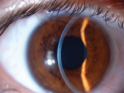 When light enters the eye; it initially encounters the tear film. The tear film coats the cornea, the crystal clear window of the eye. Inadequate tear production can cause dry eye. Located behind the cornea is the anterior chamber, which is filled with fluid called the aqueous. This clear fluid is responsible for maintaining eye pressure. Any trouble with the drainage or production of the aqueous can lead to high pressure within the eye and damage to the optic nerve, which is called glaucoma.
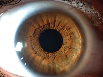 The iris is located inside the anterior chamber. The iris is responsible for eye color and acts like the aperture of a camera. It widens and narrows the pupil to enable more or less light to come into the eye. The ciliary body behind the iris is responsible for the production of aqueous fluid and maintaining eye pressure. It also contains the muscle that is responsible for focusing the natural crystalline lens.
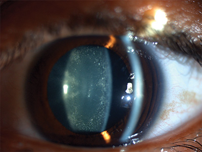 After the iris, light next traverses the natural crystalline lens. The lens focuses images of the outside world onto the retina. The lens changes shape slightly so that we can change focus when viewing objects that are near or far. As we get older, the flexibility of the lens decreases, and we become less able to change focus, particularly at near.
This condition is known as presbyopia and can be remedied with reading glasses. Ultimately, the lens loses its clarity and may turn yellow or become cloudy. This condition is called a cataract. When cataracts cause blurry vision, dull colors or other symptoms, the lens can be replaced with an artificial lens during cataract surgery.
Dr. Slakter and the team are the very best. They have been taking care of me for the last 15 years or so and it is really more than the outstanding medical specialty, which goes without saying. It is also the human care and personal consideration that makes me feel part of a team that really cares about me and all other patients equally!
MOSES D. Google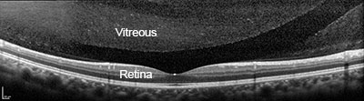
After the lens, light passes through the vitreous, the jello-like substance that fills the interior of the eye. In younger years, the vitreous is firmly attached to the retina and relatively solid. As we get older, the vitreous becomes liquefies and becomes more watery, while the solid portion shrinks. This causes it to eventually separate from the retina. In most cases, little strands or clusters of vitreous form and cast small moving shadows, which are called floaters.
Floaters are usually not a threat to vision, but they can signal a more serious condition, such as a retinal tear or detachment. If detected early, a tear or small detachment can be treated with laser in the office. Sometimes an in-office surgery involving a small gas bubble can be performed to avoid more invasive procedures. However, a retinal detachment requiring surgery in the operating room usually has an excellent chance of being successfully repaired with vitrectomy, scleral buckle, or a combination of both. Rarely, vitreous floaters are so debilitating that removal by vitrectomy is necessary.
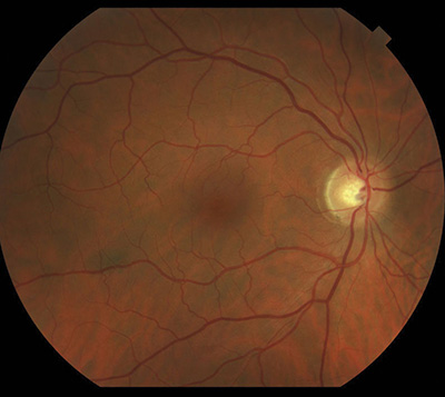 Lastly, light encounters the retina. The retina is the thin tissue that lines the innermost wall of the eye. The retina can be thought of to work much like a camera film. The retina responds to light rays by converting light into electrical signals. The optic nerve carries these signals to the brain. The outer part of the retina is responsible for peripheral vision, while the center portion called the macula is responsible for central and color vision, providing fine detail, and the ability to read.
In the center of the macula is the fovea. The fovea consists of a high concentration of special cells known as cones, making it the only part of the eye capable of perfect 20/20 vision. Any disease that affects the macula reduces central vision.
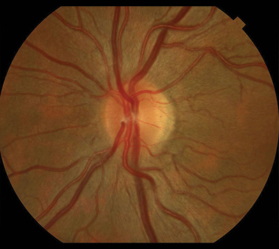
The optic nerve takes the electrical signals from the retina and transports it to the brain. The brain interprets these signals into a visual image. In glaucoma, the optic nerve becomes damaged due to elevated pressure in the eye.
Let us help you enjoy your life
Call: (212) 861-9797To Speak With An Appointment Coordinator Now