The vitreous is the clear gel that occupies about 80% of the inside of the eyeball. As we age, this jello-like substance liquefies, shrinks, and eventually separates from the optic nerve and the retina in the back of the eye. This is called posterior vitreous detachment (PVD) and occurs in most adults between 40 and 70 years of age without untoward consequences. A PVD may occur in younger individuals who have undergone cataract surgery or are very nearsighted.
When a PVD occurs, floaters are usually noticed. Floaters are small specks that move in and out of your field of vision. They may be more noticeable when looking at a plain background, such as a white page, wall, or clear sky. Some people mistake floaters for flying bugs. Flashes of light in the periphery of the visual field, particularly in low ambient light, are also a common symptom of a PVD. These flashes are a result of the retina being tugged on by the separating vitreous.
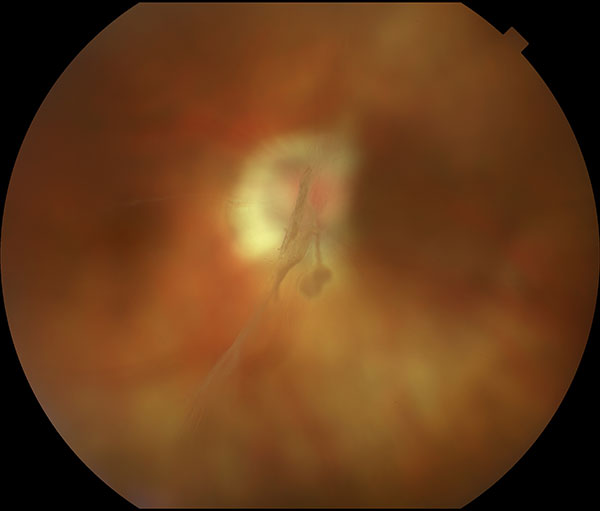
PVD is a common occurrence, which usually has no untoward consequences. However, if the retina is weak or the vitreous gel is abnormal, a retinal tear can occur, which in about 50 percent of cases eventually leads to a retinal detachment.
Being aware of the symptoms of a PVD is the first step to avoiding a retinal detachment. If you begin to notice floaters and flashers, please contact your ophthalmologist for a thorough examination immediately.
If detected early enough, retinal breaks can be treated to avoid a retinal detachment.
While early detection is the key, finding a retinal break may be difficult and requires a very thorough examination. Your ophthalmologist will dilate your pupil specifically for this exam and, using a light source and a hand-held lens, will examine the eye as slight pressure is put on the eyeball with a stick-like instrument.
This exam will bring any retinal breaks into view and show their exact size and location. In some instances, ultrasonography may be performed to help determine the status of the vitreous and if there are any retinal breaks.
Fortunately, most PVDs do not cause a retinal tear and most treated retinal tears won’t lead to a retinal detachment. However, if a retinal tear is associated with a PVD and not treated with laser, a retinal detachment is likely and further treatment will be necessary.
Dr Sorenson and personnel at vrmny are very professional and caring. I could not be more satisfied with their attentiveness.i have patronized vrmny for four years and have not been disappointed at any time. Even through the COVID emergency the doctors and staff maintained their professionalism. I am presently recovering from surgery performed by Dr Sorenson and have absolute confidence of full and satisfactory restoration of my sight.thank you Dr Sorenson
Floaters may be very distracting initially, but usually become less bothersome over time. In rare cases, floaters can be disabling to the point that removal of the vitreous and floaters by vitrectomy surgery has to be considered.
Floaters are usually not a threat to vision, but they can signal a more serious condition, such as a retinal tear or detachment. If detected early, a tear or small detachment can be treated with laser in the office. Sometimes an in-office surgery involving a small gas bubble can be performed to avoid more invasive procedures. However, a retinal detachment requiring surgery in the operating room usually has an excellent chance of being successfully repaired with vitrectomy, scleral buckle, or a combination of both. Rarely, vitreous floaters are so debilitating that removal by vitrectomy is necessary.
Top-Quality Eye Care. Our New York ophthalmologists and eye doctors publish more in the foremost peer-reviewed journals about the treatments for Posterior Vitreous Detachment, Flashes and Floaters than any other private or academic group in the United States. Many current concepts in retinal disease and some of the best treatments recognized worldwide were invented at our eye center.
Cutting-Edge Diagnostic Tools. While early detection is the key, finding a retinal break may be difficult and requires thorough examination. Our ophthalmologist will dilate your pupil specifically for this exam. Using ultrasonography, our doctors can identify the exact size and location of retinal breaks.
Groundbreaking Research. Our renowned retinal specialists lecture worldwide on retinal vascular disease and serve as academic leaders in the field as the most published group in foremost peer-reviewed journals in the U.S. Physicians at VRMNY are past or current editors of the American Journal of Ophthalmology and on the editorial board of numerous journals, including the International Journal of Retina and Vitreous, Retinal Cases & Brief Reports.
Reputation. Our reputation for outstanding eye care gives you access to the latest treatments and the best doctors to treat eye floaters. Our specialists are Castle Connolly Top Doctors, New York Super Doctors, are selected in the prestigious group of New York Magazine Best Doctors, one of the best physicians in the United States by “The Best Doctors in America,” and are consistently quoted by well-known eye specialists.
For more information about the latest treatments for eye floaters or VRMNY doctors, call the ophthalmology center at (212) 861-9797. To schedule an appointment with any of our physicians, visit our centers in Manhattan, Brooklyn, or Westchester.
Let us help you enjoy your life
Call: (212) 861-9797To Speak With An Appointment Coordinator Now