 (212) 861-9797
 (212) 861-9797
A detached retina threatens your vision. Without immediate treatment, your retina stops working, and that’s the key to vision in that eye. Ophthalmologists have used traditional surgical methods in the past to repair the retina, but today’s eye surgery relies on laser accuracy. The endolaser retinopexy/endoscopy is a diagnostic tool and a treatment aid to repair damage inside your eye from a variety of diseases and injuries. For retina laser surgery that can save your sight, call eye care center of Vitreous Retina Macula Consultants of New York for a consultation and treatment.
Pneumatic retinopexy (PR) surgery is an advanced eye procedure to repair rhegmatogenous retinal detachments using an expanding gas bubble. The gas bubble floats over the detached retinal area and gently pushes it back into position. Retina laser surgery uses a thermal laser to repair retinal damage, including:
A retinal detachment is a medical emergency that requires immediate treatment. Otherwise, this condition can lead to vision loss. An endolaser retinopexy is a common in-office procedure your ophthalmologist performs to prevent permanent damage to your retina.

The top eye doctors at Vitreous Retina Macula Consultants of New York (VRMNY) diagnose a detached retina using the latest medical technology, including diagnostic imaging. After the diagnosis, your doctor chooses from a variety of treatments, including retinopexy or a surgical procedure such as:
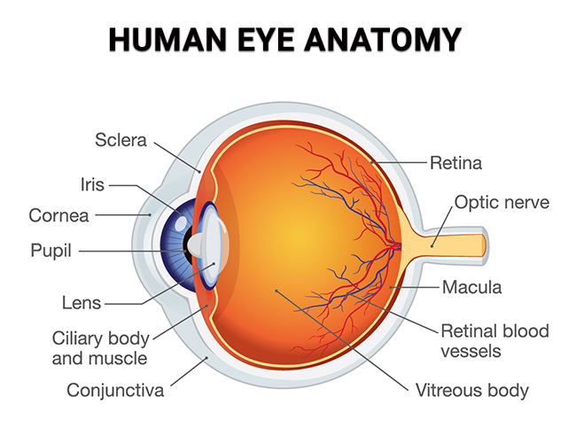
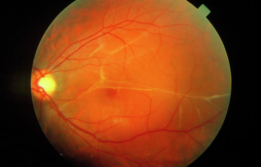
The retina is a thin membrane that lines the back part of your eyeball. The layer comprises light-sensitive cells that interpret visual images. These cells receive information and send it to the brain in real time to enable you to see. The tiny photoreceptor cells known as cones and rods help you differentiate colors, light and dark. With a retinal detachment, the supply of nourishment to this critical part of your eye is cut off.

If the retina pulls away from the blood vessels at the back of your eye, it creates an emergency situation referred to as a retinal detachment. The main types of retinal detachment are rhegmatogenous, tractional and exudative. Each form of detachment develops because of a specific situation, but each causes the retina to move from its position. Symptoms of retinal detachment include:
If you experience any of these symptoms, your retinal specialist discusses different detachment repair options, one of which is a pneumatic retinopexy procedure. To perform your surgery, your ophthalmologist uses an endolaser and an endoscopic probe. Refining the retinopexy procedure with a laser treatment and improved visualization delivers better results.
Eye specialists use the endolaser retinopexy/endoscopy procedure for improved retinal detachment repair and for a wide range of retinal complications. The advanced retina laser surgery:
During a regular eye checkup or eye treatment appointment, a top ophthalmologist diagnoses the conditions that require pneumatic retinopexy surgery. After assessing the damage to your retina, your eye doctor can opt for the advanced endolaser retinopexy/endoscopy, often combined with another procedure, to treat your retina issues.
Dr. Freund is everything one would want in a doctor: conscientious, ethical, knowledgeable, and experienced. He is ready, able and willing to explain to you your diagnosis and treatment. He’s also very personable. Highly recommended.
JOHN G. GoogleEndolaser retinopexy/endoscopy is an advanced form of retinopexy. During the procedure, your eye doctor uses an endolaser probe integrated into an endoscope fitted with a light source. The goal is to improve your doctor’s view inside your eye, which is a big problem when performing retinal detachment repair. The technique improves the outcome of retinal surgery. Some of the benefits of endolaser retinopexy/endoscopy include:
After reaching a diagnosis, your eye doctor uses state-of-the-art equipment at the VRMNY office nearest you to perform the retinopexy procedure, in combination with endolaser and an endoscopic probe. You’re always in the safest hands at this New York City-based eye practice.
The traditional retinopexy procedure forms a barrier that prevents fluid from entering through a retinal tear or hole. The endolaser retinopexy/endoscopy procedure is more advanced, as your eye doctor also uses an endolaser probe with an endoscope. The main steps in the retinopexy procedure include:
Endolaser retinopexy/endoscopy requires specialized skills that the ophthalmologists at VRMNY possess. These retina specialists and surgeons are renowned medical researchers and innovators in eye treatments. Contact a top eye specialist at Vitreous Retina Macula Consultants of New York today to protect or restore your vision.
Let us help you enjoy your life
Call: (212) 861-9797To Speak With An Appointment Coordinator Now