Today, eye surgeries make use of the latest equipment, techniques and knowledge. They’re mostly in-office, outpatient procedures with a minimal amount of recovery time. And when you rely on the best retina doctors at Vitreous Retina Macula Consultants of New York, you can be sure you get the most appropriate procedure done properly. Call eye doctors today if you’re having vision problems or eye pain.
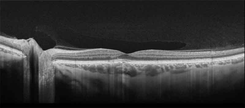
Optical Coherence Tomography (OCT) uses a non-invasive safe beam of light with special processing to look at the structures within the retina in very high detail. The anatomic features visible with OCT are much more detailed than that seen by normal examination. Seeing the retina from this cross sectional viewpoint provides a powerful way to detect, diagnose, and treat retinal disease. Pathological changes associated with age-related macular degeneration, diabetic macular edema, central serous chorioretinopathy, and retinal edema associated with retinal vein occlusions can be detected and monitored with this form of imaging. OCT imaging is crucial in diagnosing and following glaucoma with scans designed to calculate changes to the optic nerve and surrounding nerve fiber layer. OCT is also imperative in measuring deeper layers, such as the choroid, which can be affected in many ocular and systemic diseases.
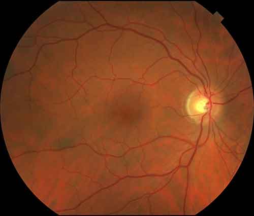
Fundus Photography is a term used to describe taking a picture of the back of the eye, an area more specifically called the retina. This allows the physician to document any present pathology as well as track the progress of retinal disease over time. With use of different filters at specific wavelengths, certain parts of the retina can be isolated for ideal perspectives. By using special light filters we can look at molecules that accumulate in health and disease in a process called autofluorescence imaging. By imaging the amount of these molecules we can learn about the functioning of the cells in the eye.
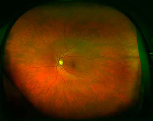
Until recently it was difficult to photographically evaluate anything other than the back of the eye. However retinal changes can sometimes extend beyond what can be visualized with a standard fundus camera. In these cases, the best option may be to document the pathology with an ultra widefield camera. At VRMNY, we have multiple wide angle camera platforms that are capable of capturing many times the area of a standard camera can photograph. This has revolutionized the diagnosis, treatment, and monitoring of conditions of disease affecting the periphery of the retina, ranging from vascular disorders to retinal detachments and tumors. It has also greatly enhanced patient education, as features of the disease that were once only visible to the examining physician can now be shown to the patient on a high-resolution screen.
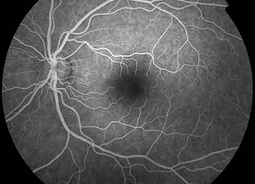
Angiography is a procedure used to view circulation and leaking or damaged blood vessels in the retina, which commonly occur in diseases such as diabetic retinopathy, vascular occlusions, age-related macular degeneration, central serous chorioretinopathy, and hypertensive retinopathy. Fluorescein angiography, which uses a bright yellow contrast dye, shows the circulation of the retinal vessels, while Indocyanine Green angiography uses a green-colored dye that highlights the choroid, a layer deeper than the retina. The dye is injected into the arm, and a rapid sequence of pictures are taken to view the dye as it flows into the eye. The test only takes a few minutes and provides vital information for the physician to decide the best course of treatment.
One of the most professional, knowledgeable and yet friendly team of doctors and professionals in NYC. I have never had my eyes examined so thoroughly and feel so much better knowing that my eyes are in good hands. Dr Klein himself attends to almost all patients and the level of attention and care is phenomenal.
ROBIN G. Google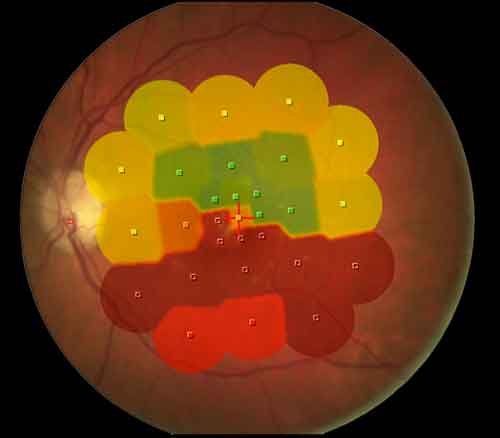
Assessing visual acuity with only the Snellen Chart commonly misses significant problems sparing the very central portion of a patient’s vision. Microperimetry can precisely map areas of decreased retinal sensitivity and pinpoint the cause of visual problems. This can then be correlated with other imaging modalities, such as angiography, OCT, or Adaptive Optics to provide the physician with a complete picture of your retinal condition.
Because of our recognized status, many manufacturers utilize our department to develop and test new imaging equipment before it’s available on the open market. We pride ourselves on having access to a wide variety of advanced prototype imaging technology not generally available to other practices to provide our patients with the most cutting-edge care.
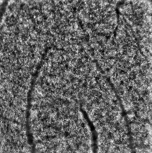
A technology called Adaptive Optics is able to eliminate ocular aberrations that distort and diminish retinal image quality, making it possible to visualize individual retinal cells of the photoreceptor layer in the retina. The ability to see retinal disease on a cell-by-cell basis allows a more precise study of the progression of disease or response to treatment, such as with drug toxicity, retinal degeneration, and macular hole and detachment repair.
Swept Source Optical Coherence Tomography is a type of research OCT that utilizes rapidly tuned frequencies of light over a broad bandwidth, allowing very fast, deep, and wide scanning of the retina and adjacent tissues with good penetration through cataracts. This provides a more detailed cross sectional view of the eye, improving recognition of structures that are unable to be seen on conventional OCT, such as conditions that affect the choroid and vitreoretinal interface. These conditions can include choroidal tumors, central serous chorioretinopathy, vitreomacular traction, and proliferative diabetic retinopathy complicated by epiretinal membrane formation and tractional detachment.
OCT Angiography (OCTA) is a scan that detects the motion of flowing blood cells. OCTA creates an image similar intravenous angiography but without the injection of contrast dye, making this a non-invasive alternative to the traditional procedure. We anticipate this form of imaging to be the next big wave in ocular imaging and VRMNY physicians are at the forefront of this new technology.
For more information about the technologies we use in the center for ophthalmology in New York City, visit one of our locations or call Vitreous Retina Macular Consultants of New York (VRMNY).
Let us help you enjoy your life
Call: (212) 861-9797To Speak With An Appointment Coordinator Now